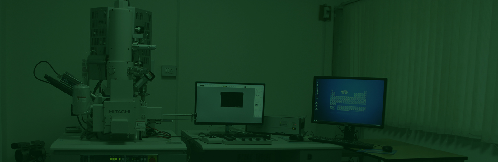
Manufactured by Hitachi Ltd, Japan With Power backup, computing and Imaging facilities
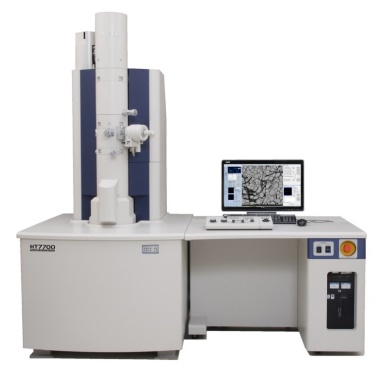
Manufactured by Hitachi Ltd, Japan With Power backup, computing and Imaging facilities
ICAR/SAU/SHU/Govt R&D
EM sample observation: Rs 1250 + 18% GST per image
Block preparation and EM: Rs 6000 + 18% GST per image
Pvt/Industries
EM sample observation: Rs 4000 + 18% GST per image
Block preparation and EM: Rs 10000 + 18% GST per image
Dr. D K Samuel Principal Scientist, Division of Plant Protection, Email: duleep.samuel@icar.gov.in , Phone +91-80-23086100 Extn: 306
Magnification: ×50 - ×1,000, (low Mag mode) ×200 - ×200,000 (High Controst mode), ×4,000
- ×600,000 (High Resolution mode)
Accelerating voltage: 40 - 120 kV (100 V/step variable)
Image output: : 8 M pixel Main Camera
640 × 480/ 1,280 × 960/ 2,560 × 1,920/ 5,120 × 3,840 pixel,TIFF (8 or 16 bit) , BMP, JPEG images. With auto nano / micron marker
Auto focus. Microtrace, Autodrive, Autophoto, features Auto-gun alignment, Live FFT display, Measurement function, Low dose and auto pre-irradiation facility
Image output in CD/DVD
The Transmission Electron microscope can image viruses and other plant pathogens. It can also image metallic nano particles. upto 300000 times primary magnification This Variable Pressure Scanning Electron Microscope can be used to image structure of bio materials upto 60000 times primary magnification
Sample dimensions, Size distribution.
The sap neds to be extracted and applied on TEm grids, After addition of PhosphoTungstic Acid or Uranyl Acetate stain, The sample needs to be washed and dried. This can be imged within a day.
The sample needs to be fixed with Osmium tetroxide or Glutaraldehyde or Paraformaldehyde and then needs to be dehydrated in an alcohol series. Followed by Embeding in Epoxy resin blocks, The cured blocks would then be cut in a Ultramicrotome using glass blades. The cut sections ned to be in 40 – 50 na1nometers (nm) thick, The sections would need to be stained and imaged after drying. Time requirement will be 2 weeks.
Nano / Miro structure, morphological